Jess Austin

Understanding Equine Gastric Ulcer Syndrome (EGUS): A Guide for Horse Owners

Understanding Equine Gastric Ulcer Syndrome (EGUS): A Guide for Horse Owners

Gastric ulcers are a widespread issue in horses, particularly among performance, sport, and even leisure horses. Equine Gastric Ulcer Syndrome (EGUS) occurs when the lining of the horse’s stomach is damaged by excessive acid exposure, leading to pain, poor performance, and behavioural changes.

Studies show that EGUS affects:

  • 80–100% of racehorses 
  • 93% of endurance horses during competition
  • 58% of show and sport horses
  • 11% of leisure horses
  • Up to 57% of foals – with risk rising to 97% after weaning

Given these statistics, it’s crucial for horse owners to understand the signs, causes, and treatments of this common but often overlooked condition.

What Causes Gastric Ulcers in Horses?

Unlike humans, horses produce stomach acid continuously—even when not eating. When their stomachs are empty for extended periods, the unbuffered acid can damage the stomach lining, especially the squamous (upper) region.

Key risk factors include:

  • Fasting & Limited Forage: Long gaps between meals allow acid to build up.
  • High-Grain Diets: Increase acid production and reduce protective saliva.
  • Intense Exercise: Causes acid to splash upward, irritating the stomach lining.
  • Stress: Travel, competition, stall rest, and routine changes all contribute.
  • NSAID Use: Medications like bute, Danilon, or Fynadine can reduce natural defenses.
  • Limited Turnout: Horses kept in stalls are more prone to ulcers than those on pasture.

Recognising the Signs

Symptoms of gastric ulcers can be subtle and are often mistaken for behavioral or training issues. Some horses show no outward signs, even with severe ulcers.

Look out for:

  • Decreased appetite, especially for grain or concentrates
  • Weight loss or poor body condition despite normal intake
  • Behavioural changes such as girthiness, bucking, or resistance to work
  • Dull coat
  • Mild or recurrent colic especially after eating
  • Excessive salivation or teeth grinding, often misattributed to dental problems

Diagnosing EGUS

The most reliable way to diagnose EGUS is through gastroscopy – a procedure where a camera is passed into the stomach to inspect for ulcers directly. Prior to the procedure, horses must be fasted (no food for 12 hours, no water for 4 hours) to ensure clear visibility.

Treating Gastric Ulcers

The goal of treatment is to reduce stomach acid and support healing, often through a combination of medication and lifestyle management.

Treatment options include:

  • Omeprazole (Gastrogard/Peptizole): The only UK-licensed treatment; reduces acid production. Typically given once daily for 4 weeks, followed by a tapering dose.
  • Sucralfate: Coats and protects the stomach lining, often used alongside omeprazole.

Nutritional adjustments:

  • Provide constant access to hay or pasture
  • Reduce high-starch and grain feeds
  • Offer small, frequent meals
  • Use slow feeders if ad lib hay isn’t feasible
  • Feed a small amount of alfalfa 30 minutes before work to reduce acid splash

Stress management:

Maximise turnout, reduce travel, and adjust training routines.

Preventative treatment:

  • Consider omeprazole during times of high stress or competition.

Most horses begin to feel better within a few days of starting treatment, but full healing can take 3-4 weeks or more, depending on severity. A follow-up gastroscopy is recommended to ensure ulcers have resolved.

How We Can Help

At Shotter & Byers, we offer gastroscopy services both at a facility we work closely with based in Epsom or at your own yard.

Whether you’re concerned about your horse’s behaviour, performance, or just want peace of mind, our team is here to support you every step of the way.

📞 **Get in touch today to schedule a consultation or gastroscopy appointment. **

Understanding Equine Gastric Ulcer Syndrome (EGUS): A Guide for Horse Owners Read More »

Equine Flu Vaccination Changes from 1st of January 2024

We want to inform you that the vaccination rules for equine influenza have recently been updated in accordance with advice from British Equestrian. Please be aware that these rules may vary between clinics.

We want to ensure that your beloved horses and ponies vaccines are up to date based on the latest guidelines. Please don’t hesitate to contact us if you have any queries!

Equine Flu Vaccination Changes from 1st of January 2024 Read More »

EQUINE HERPES VIRUS

What is equine herpesvirus (EHV)?

EHV stands for equine herpes virus. The two most significant types are Equine Herpes Virus 1 (EHV-1) and Equine Herpes Virus 4 (EHV-4).

Respiratory EHV is endemic in the UK, which means it is everywhere however clinical signs are often mild, and your horse may even have had it in the past without you realising it. The respiratory form of the disease can be caused by both EHV-1 and EHV4: it is reported that 80-90% of horses are infected with EHV before the age of 2 years old.

The neurological form of the disease is rare and obviously is very serious and can be fatal. This form is usually due to EHV-1.

EHV can also be associated with abortion, still birth and severe neonatal illness.

Some horses are life-long carriers, and the virus can be re-activated in these individuals and cause clinical signs and spread to others. Risk factors for re-activation and consequent spread include transport, strenuous exercise, and at equine events.

Most spread of the virus occurs by close horse-to-horse contact, but it can also be spread by sharing equipment such as tack and mucking out tools.

Should you be vaccinating?

The EHV vaccination should not be considered as an alternative to good biosecurity. We strongly encourage horse owners to quarantine all horses newly arrived on their premises.

It is recommended that the following groups of horses should be vaccinated:

·         Broodmares should receive their EHV vaccinations every year.

·         Racehorses are generally considered high risk and, depending on the jurisdictions to which they are travelling, vaccination may be obligatory.

·         Horses which travel away for competitions, particularly when this involves overnight stays in shared stabling.

·         Horses which are living on yards where other horses travel away frequently

It is important to remember however that whilst the EHV vaccination reduces shedding of the virus and makes clinical signs milder, it does not necessarily abolish either. However, reducing shedding helps to minimise risk to other horses when a horse is infected with EHV.

There is currently no evidence that vaccination prevents the development of the neurological form of the disease.

To provide effective immunity against respiratory and neurological disease caused by EHV 1 and EHV- 4 a primary course of 2 vaccinations should be given followed by a booster vaccination every 6 months.


  • 1st vaccination: Can be given to any horse over the age of 5 months.
  • 2nd vaccination: To be given 4-6 weeks after the 1st vaccination.
  • 6-month booster: To be given within 6 calendar months of the 2nd vaccination.


To provide effective immunity against abortion caused by EHV 1 and EHV-4 a course of three vaccinations should be given to a mare during her 5th, 7th and 9th months of pregnancy.


To book in your vaccinations please contact Shotter & Byers on 01306 627706.

What is equine herpesvirus (EHV)? Read More »

CCI2 Winner Team Breach Eventing

Winner Winner – Congratulations to Fiona Breach Eventing

Congratulations to our lovely client, Fiona at Team Breach Eventing who won the CCI2* at Ballindenisk International Horse Trials on her dressage score of 45.6

The other highlight of the week was the donkey derby in which Georgia Bale Eventer and Marieke both participated without much success but having had a fantastic time stretching their equestrian skills to the max.

Marieke was also the FEI treating veterinarian for the event.

Winner Winner – Congratulations to Fiona Breach Eventing Read More »

Penetrating wounds of the horse’s hoof

Penetrating Wounds of the Foot

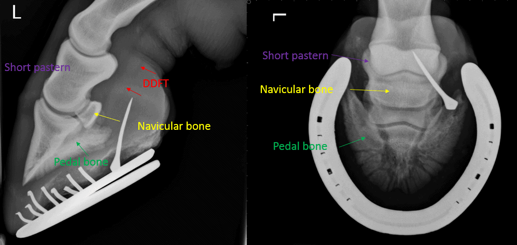
Penetrating wounds of the horse’s hoof are quite common with farrier nails and joinery screws being the most frequent cause. When a sharp object has penetrated the horse’s foot, it can potentially cause damage to the sensitive tissues and structures inside. Depending on the depth and location of entry, the penetration can result in very serious damage, and so a penetrating foot injury should always be carefully assessed by a vet.

The equine foot is comprised of hoof wall, sole and frog. Once the hoof wall or sole has been penetrated, the consequences will depend on what structures are damaged. The frog is the softest part of the foot and is prone to penetrating injuries. As the frog has deep grooves (or sulci) on either side, sharp objects can become wedged, before potentially being driven further into the foot.

The horse’s foot contains two very important synovial structures, the coffin joint and the navicular bursa. If either of these structures are penetrated, potentially life-threatening synovial bacterial infection almost always occurs. Prompt assessment of penetrating foot wounds should therefore be performed to assess the site, depth and angle of penetration.

The navicular bursa, is only about 3 cm from the sole; in a pony it is even less. Therefore, a short nail does not have to penetrate very far to reach the bursa. If bacterial infection develops in the navicular bursa or the coffin joint (or both) then there can be disastrous consequences. The body’s immune system, in its attempt to eradicate the infection, also damages the synovial structures themselves, for example destroying the smooth surface of cartilage. In a matter of just a few days, the combination of bacterial infection and inflammatory response can cause such extensive damage to a synovial structure that a horse will be lame for the rest of its life, even with treatment, and in such situations the horse often needs to be put to sleep.

If a nail extends deeper into the foot, it can come into contact with the pedal bone. If there is sufficient force of penetration, the pedal bone is damaged and becomes infected (septic pedal osteitis). Bacterial infection can “eat away” at the bone causing serious problems and treatment is not straightforward, as antibiotics find it very difficult to penetrate infected bone. Usually the entire puncture wound tract must be pared away and the pedal bone scraped away back to healthy bone. Obviously, this procedure has a long recovery time as healthy tissue must repair the hole made in the foot.

Tetanus from any penetrating foot injury is a potentially fatal disease that is extremely difficult to treat, however it is preventable by vaccination (see Vaccination page)

In most situations it is better to leave the nail in place, as long as the horse is not standing on it, driving it deeper into the foot. Try to keep the horse calm, hold the foot up if necessary, and call out one of our vets with an xray machine, as an emergency.

If the nail has only penetrated a short distance into the soft tissues of the sole, the nail is removed, and the entry tract is pared out and enlarged to ensure any pus can drain from the infected site. The tract is drained with iodine, and possibly antibiotic saline and a poultice is placed, to keep the wound clean and draining. Tetanus cover might be given if needed, and possibly antibiotics.

If deeper structures are suspected to be involved, our vet may decide to take radiographs before they remove the nail. This will tell them exactly where the nail has gone and which structures have been damaged by the nail. Bony damage may need surgical treatment and prolonged antibiotics. If the navicular bursa or coffin joint are shown to have been penetrated, the horse will need to be referred as soon as possible to an Equine Hospital for possible further MRI of the foot, and to surgically flush out the synovial structure.

Penetrating Wounds of the Foot Read More »

horse jumping in school

Exporting Your Horse

Whether you are off for a race in Dubai, eventing in Kentucky or simply moving home to France, we can help you every step of the way with our helpful guide of what you will need to do.

First of all you need to consider whether you will be flying, using the train or transporting your horse by water and contact some transportation companies for quotes.  They should also be able to offer additional guidance on procedures and preparation.  Also check your horse insurance will cover the trip and that your transporter is properly covered.

If competing, check with your competition federation such as the FEI on rules surrounding vaccines and immunizations and permitted drugs as well as the need for quarantine which varies from country to country.

Your destination country’s government website should also hold some helpful information on what documentation your horse will need to have traveling with it and when it arrives.

Always keep in mind timings, these can be very strict and difficult to adhere to if you do not prepare well in advance. Some vaccines need to be administered months in advance for example and quarantine can also be time consuming and costly.  If you are competing you will need to also think of your horses fitness and recovery time.

For Travel within the EU all that is needed is an intra trade certificate that needs to be applied for through DEFRA – https://www.gov.uk/guidance/export-horses-and-ponies-special-rules – they then notify us and we come out and do the health certificate within 48 hours of travel.

For travel outside of the EU every country is different but a lot more notice is often needed as vaccinations, bloods and/or swabs and other treatments are needed with a specific time frame – your export company should be able to liaise with us and arrange these for you to ensure it is done correctly as even if your horse is aloud out of the UK without the correct testing and documentation it may not be allowed into its destination.

If you are unsure of the veterinary requirements, why not give us a call and see if we can assist.  We often assist with the transportation of horses around the world and would be happy to help.

Exporting Your Horse Read More »

Extracorporeal Shock Wave Therapy (ESWT)

Worming

Controlling Parasites in your Horses

There are three groups of internal parasite which can damage the gut and cause disease in the horse. The information below will hopefully explain the importance of developing a good worming regime, using the appropriate products at the correct time:

ROUNDWORMS are grazed of f the pasture as microscopic larvae which bury their way through the gut and over many months migrate around certain organs of the body. They then return in the larval form to the wall of the intestine where they live for a while before bursting out into the gut to form adult worms which produce eggs to contaminate your pasture.

TAPEWORMS have an interesting life cycle which cannot be completed without an ‘intermediate host’ known as the harvest mite found on the pasture during the summer and autumn and to a lesser degree in hay. When the horse ingests the mite, the tapeworm is liberated in an immature form and over many weeks, changes into an adult tapeworm.

BOTS are a bee-like fly. Eggs are laid by the fly on the horse, mainly on the legs, which are ingested when the horse’s mouth comes into contact with the eggs. The eggs hatch by this process, the larvae bury their way through the back of the tongue and all the way down to the stomach, where they erupt and form masses of large grubs which cling to the stomach wall.

There are many different horse worming products on the market. We believe that EQUEST & EQUEST PRAMOX will provide the spectrum of ant parasitic cover necessary to protect your horse’s health.

Your worm control will only be as good as your management. Using the appropriate wormer, picking up droppings in the field, rotating your grazing fields, and ploughing, liming and reseeding every few years will help prevent worm infestation on the pasture. Any new horse introduced to a yard should be wormed with EQUEST and kept in a box or restricted paddock for a couple of days with collection and disposal of droppings prior to any introduction to communally grazed pasture. The following worming protocol is advised:

  • Winter/Summer: EQUEST (Adult and encysted roundworms / Bots)
  • Autumn/Spring: EQUEST PRAMOX (Adult and encysted roundworms / Bots/Tapeworms)

Remember to either keep the horse in for 24hours after worming or to make sure that ALL droppings are picked up ASAP. No worm product kills 100% of worms, hence the necessity to follow the above protocol.

Worming Read More »

Chronic grass sickness

Grass Sickness

Clinical signs are attributable mainly to stasis of the entire alimentary tract, and include depression, inability to swallow, lack of appetite, gastrointestinal distension and impaction, abdominal pain, sweating, elevated heart rate, muscular tremors, weight loss and drooping eyelids. There is no specific treatment for the disease. The majority of affected horses are euthanised on humane grounds, but some horses with mild chronic grass sickness may survive with intensive nursing care.

Acute Grass Sickness

This is the most serious. It is of rapid onset, the horse is severely distressed, has an elevated heart rate, and patchy sweating, may be dull and depressed. Moderate quantities of green fluid may be produced from the nostrils (this should always be considered a serious signs in a horse). There is lack of gut movements, and usually no faeces will be produced. There are often muscle twitches involving the muscle of both fore legs. Horses with acute grass sickness are often confused with colic, but rarely roll or go down. Veterinary attention is required urgently. Often when a stomach tube is passed a vast quantity of grass fluid will flow back. Destruction on humane grounds is the only option.

Subacute Grass Sickness

Affected animals rarely have gastric reflux (green discharge) but will show most of the signs displayed by the acute cases. An inability to swallow is a prominent sign along with drooping of the upper eyelid. Subacute cases are always fatal; the course of the disease is 2 – 7 days.

Chronic grass sickness. These cases are less severely affected than subacute cases and may be able to eat small amounts of food. Some of these horses may survive for considerable periods of time and may respond to treatment.

However many are left permanently damaged, and unfortunately euthanasia is the common outcome. Currently there is no way of treating this disease.

Risk factors include:

  1. Recently introduced to new pasture.
  2. Previous history of grass sickness on pasture.
  3. No hay or conserved feed being fed.
  4. Aged 2 to 7 years.
  5. Highest incidence April – July (but can occur all year
    around).

It must be stated however that all horses can be affected by grass sickness. Grass sickness is caused by widespread severe damage to the neurons (nerves) in the autonomic nervous system (this is the part of the nervous system that helps control the intestines).

Patchy sweating is common in all forms of grass sickness.

It is not possible to protect your horse 100% from grass sickness. The following advice may help:

  1. Always feed some conserved forage (hay) even when horses are turned out 24 hours a day.
  2. Don’t put horses in the at risk age group (2-7 years) out onto pasture where there has been previous cases.
  3. Make sure your horse is healthy with regular worming or fecal egg count monitoring.

The cause of grass sickness has remained a mystery for the last ninety years. Research workers have recently made some interesting advances, but there is still much left to find out about
grass sickness. We need to raise awareness and we need more funding for research.

Grass Sickness Read More »

baby horse sitting

Foal Septicaemia

Septicaemia is one of the most serious conditions in foals, and unfortunately a relatively common occurrence in neonates. It is caused by infection of the bloodstream which causes inflammation all over the body. As soon as a foal is born it is exposed to bacteria. Two of the most vulnerable areas for bacteria to enter a newborn’s system are through its navel area and through its mouth. Therefore it is vital that two things occur shortly after birth: the navel is disinfected with a gentle iodine and the foal must receive the mare’s first milk (colostrum). It is from the mare’s colostrum that the newborn receives vital antibody protection against bacteria.

It is extremely important to observe newborn foals in the first 24 hours as this is when symptoms of Septicaemia will often show up. An unhealthy foal will go downhill very quickly and without veterinary care may die in a matter of hours.

A healthy foal will be exploring it’s surrounding, not be shy about trying out its legs, take frequent naps but be up and alert again after that. The septic foal will just slowly decline, want to sleep all the time, and become less responsive to stimulation.

There are a lot of different clinical signs that can be associated with Septicaemia. Most affected foals will have several of these signs, but not necessarily all of them. Some of these signs can also be caused by other problems, but remember that a newborn foal with problems of virtually any kind is at higher risk for developing Septicaemia. Signs of Septicaemia may include:

  • Depression
  • Lack of suckle reflex (normal foals should try to suck on a person’s fingers or a bottle nipple if placed in the foal’s mouth)
  • Fever (too high a temperature), or hypothermia (too low a temperature)
  • High heart rate (most new born foals have a heart rate between 80-120 beats per minute)
  • High respiratory rate or trouble breathing
  • Gums and lips an abnormal colour (e.g. dark red or purplish)
  • Swollen, painful joint(s)
  • Cloudy eyes (i.e. anterior uveitis)
  • Seizure activity if the brain is inflamed
  • Lack of urine production or renal failure

In most cases, the appropriate action to take if you have a foal with any combination of these signs is to call your veterinarian as soon as possible. A sick neonatal foal is an emergency.

Foal Septicaemia Read More »

Referral Veterinary Services

Sarcoids

Sarcoids are the most common form of equine skin tumour, they are classified as low grade fibrosarcomas (tumours). Although common, sarcoids vary greatly in appearance and size and the nature in which they grow and the response to treatment. It is this variability which makes them such a challenge for owners and vets to manage and treat. Certain breeds are more commonly affected with thoroughbreds being over represented. Geldings appear to be more commonly affected than mares. Although a tumour, sarcoids do not spread internally. Sarcoids may rise at any site on the skin but most commonly at sites where flies land; chest, groin, sheath, belly and around the face as well as at sites of previous or current wounds. The majority of cases arise between the years 3 to 6 of age, however they may appear later in life. It is thought that flies play a role in the spread of sarcoids between horses.

There are various types:

Occult

Appear as hairless grey scaly , rough circular patches of mildly thickened skin. They are the least distinctive and often mistaken for rubs or ringworm.

Verrucous (warty) sarcoids

Wart like in their appearance and often greyish in colour. They are usually slow growing and not aggressive however any interference or trauma may result in these lesions changing to a more aggressive form of sarcoid. They are commonly found around the face, armpit and sheath regions of the body. They may appear singularly or coalesce into groups forming larger lesions.

Nodular sarcoids

Firm and nodular in nature these sarcoids are more common in the eyelid, armpit, inside thigh and groin regions. Often the nodules are freely mobile under the skin although they may be attached to overlying skin. The overlying skin is usually normal but may start to thin and ulcerate. They may remain static in size for many years but can become aggressive if interfered.

Fibroblastic

Fibroblastic sarcoids are often aggressive and have a fleshy granulomatous appearance.  Sometimes they are pedunculated but can also be firmly attached.  Fibroblastic sarcoids can also develop on wounds and may closely resemble proud flesh.

Malevolent

This is the most aggressive form of sarcoid and most commonly affects the face, elbow and inside thigh regions of the body. Can spread over a wider area and quickly grow in size. They appear like ulcerated nodules but tending to group into larger bundles. This form of sarcoid is more difficult to treat but is rarer.

There may be lesions/sarcoids that display qualities of two or more sarcoid types. It is important to note that no two sarcoids are the same and treatment and response to treatment may vary between sarcoids.

Treatment

There are a range of treatment options available, dependent on type of tumor, location of the tumor and your budget.  Common treatment methods include the use of creams such as Liverpool Cream, the use of a ring around the tumour, supplements and / or laser treatment and / or a range of alternative therapies.

If you think your horse may be suffering from sarcoids, please give the practice a call so that we can review and consider if treatment is recommended.

Sarcoids Read More »