25 May 2017

Equine metabolic syndrome

Equine Metabolic Syndrome

What is Equine Metabolic Syndrome

Equine metabolic syndrome can be defined as a collection of risk factors that are associated with an increased susceptibility to laminitis. Characteristically these are obesity, patchy accumulation of fat, laminitis and insulin resistance.

Diagnosis of Insulin resistance is difficult because insulin levels are affected by a huge number of factors including diet, exercise, pain, stress, illness or Cushings. The gold standard method is not practical for regular use but there are a number of other methods available.  The most commonly used for practical reasons is a single blood sample taken after an overnight fast to test for high resting insulin (Resting hyperinsulinaemia).

However, a normal or low resting insulin does not necessarily rule out insulin resistance.  If a horse is displaying a number of other signs of EMS a glucose challenge test can be performed. A measured amount of glucose is fed following a 12 hour fast and a blood sample is taken 2 hours later.  Horses with IR are likely to have an excessively high insulin level and/or a delayed return to normal blood glucose levels.

If EMS is identified in your horse it is very manageable with dietry changes with or without the addition of medicines such as levothyroxine sodium or metformin to increase insulin sensitivity.

The goals of treatment and management are:

  • Induce weight loss in obese horses
  • Improve insulin sensitivity through weight loss, diet and exercise
  • Avoid dietary triggers for laminitis

Equine Metabolic Syndrome Read More »

x-rays scanning

Digital Radiography – X-RAYS

Digital Radiography

At Shotter & Byers we are proud to own five top of the line wireless Eklin digital x-ray machines.  These allow us to take exceptional quality images that can be viewed instantly beside your horse without the constraints of wires.

We also have a wired system that displays the images instantly and a number of x-ray generators to be used with more traditional digital x-ray plates that are developed at our office.

This array of x-ray equipment allows us to provide you and your horse with an exceptional and fast veterinary diagnostic service.

Digital Radiography – X-RAYS Read More »

Diagnosing Lameness

Diagnosing Lameness

All cases of lames are different but this post gives a general breakdown of some of the steps that may go into a lameness workup.

  1. Review of Medical History

Your vet will ask you questions about your horse and gather any information they may feel is relevant to the current situation.

  1. Visual Examination at Rest

By visually examining your horse at rest your vet can note its conformation, balance, weight-bearing and look for any signs of injury.

  1. Examination in Motion

Your vet may wish to see your horse in motion.  It may be required to be seen moving in a straight line, in circles, in hand or on the lunge. Occasionally your vet may also request to see your horse under saddle to get a more compete view of the presenting lameness.

  1. Hands on Exam

Your vet will palpate your horse, checking joints, bones, muscles, tendons and ligaments for any signs of injury or abnormality.

  1. Application of Hoof Testers

This piece of equipment allows your vet to apply pressure to areas of the foot to look for abnormal sensitivity or pain.

  1. Flexion Tests

Your vet will hold a leg in a flexed position for a period of time before evaluating your horse in motion once more. Your horses’ response to flexion tests can help identify the cause of the lameness.

  1. Nerve and Joint Blocks

Local anaesthetic can be injected into joints or around nerves to numb certain areas.  Blocking is a very useful diagnostic technique for identifying the location of a lameness.

  1. Imaging

Imaging is a further step towards identifying the cause of lameness. There are two main imaging techniques that can be brought to your horse, x-ray for bony structures and ultrasound for soft tissues. Other methods such as MRI, CT and scintigraphy are available but often require sending your horse to a hospital facility.

We offer state of the art digital x-rays which can be viewed instantly on a computer beside your horse and diagnostic ultrasound.

Diagnosing Lameness Read More »

Chronic grass sickness

Colic

Colic

Most horses will suffer from colic at some point in their lives. There are a huge number of possible causes for colic and they vary greatly in severity however they can all share similar symptoms.  It is very important that you are able to recognise the signs of colic so your horse can receive the appropriate medical care as soon as possible.

What is colic?

Colic itself is not a disease but is a term used to describe abdominal (belly) pain in horses.

Signs of mild colic

  • Pawing or scraping the ground.
  • Turning the head to look at the abdomen (‘flank watching’).
  • Kicking or biting at the abdomen.
  • Stretching out as if needing to urinate.
  • Restlessness – getting up and down frequently

Signs of severe colic

  • Rolling
  • Lying on its back.
  • Recumbencey (unable to stand)
  • Increased respiration rate
  • Increased heart rate
  • Red/purple colour of mucous membranes (gums and eyes)

What causes colic?

There are many causes of colic and it is often impossible to pinpoint the exact cause. However, there are a few risk factors:

  • Change in diet
  • Change in management
  • Change in exercise
  • Heavy worm burden

What should I do if I think my horse has colic?

  • Remove any feed from your horse and contact us for advice.
  • If you are able to, take your horses temperature, heart rate and respiration rate.
  • If your horse is rolling, keep your distance and stay away from harm. When possible remove any objects that may injure your horse while rolling.
  • If the colic is mild and your horse is calm it is acceptable for them to lay down quietly in their stable.
  • In some cases hand walking your horse may ease mild colic signs and reduce their urge to roll.
  • Your horse may become more violent and distressed, desperately wanting to go down and roll. In this case it may be safer for you and them to let them roll in a well bedded stable than it would be for them to go down while walking in the yard.
  • Do not give your horse any medication unless instructed to by your vet. Some drugs, particularly finadyne, can mask colic signs and prevent an accurate diagnosis.

Colic Read More »

choke white horse

Choke – Oesophageal Obstruction

Choke – oesophageal obstruction

What is choke?

The term choke can be misleading. Choke in horses refers to an obstruction of the oesophagus, not the trachea as in humans.  This means while choking your horse will be able to breathe, despite being in distress.

What causes choke?

  • Poor dental health
  • Horses with dental abnormalities such as missing teeth, malocclusions or sharp points are unable to chew effectively. This puts them at a far higher risk of choking compared to those that have good dental health.
  • Bolting feed
  • Some horses eat too fast and do not chew properly leading to choke. This can be avoided by placing large stones in with their hard feed which they must eat around, slowing them down.
  • Dry food
  • Dry food is often harder for your horse to swallow than damp or wet food. This cause of choke is easily avoided by slightly wetting your horses feed. This is very important for sugar beet which must be properly soaked before feeding.
  • Foreign objects
  • Occasionally your horse may eat something it shouldn’t, such as a piece of wood, which could get stuck in their oesophagus

Signs of choke:

  • Distress
  • Coughing
  • Not interested in food.
  • Difficulty swallowing.
  • Head and neck extended in a downwards position.
  • Nasal discharge
  • Saliva drooling from the mouth

What to do if your horse is choking

You should remove any remaining food from your horse and call us for advice.

Your vet will likely sedate your horse and give them some anti-inflammatory medication.  This will help your horse relax and may release the blockage.

Some obstructions require your vet to pass a stomach tube through your horse’s nose. This allows them to lavage the blockage with water to remove it through the tube, this can be a time consuming process if the blockage is large.


Choke – Oesophageal Obstruction Read More »

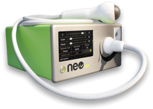
Extracorporeal Shock Wave Therapy (ESWT)

Extracorporeal Shock Wave Therapy (ESWT)

Extracorporeal Shock Wave Therapy (ESWT)

ESWT has been used for many years in human medicine to treat many orthopaedic conditions such as tennis elbow, shoulder and heel injuries etc and is now a commonly used and effective treatment in equine medicine.

Some of the conditions commonly treated using ESWT include:

  • Osteoarthritis
  • Wounds and Chronic Wound Care
  • Acupressure Points
  • Non-union or delayed-healing of Fractured Bones
  • Joints
  • Bone
  • Large Muscles
  • Tendon Injuries
  • Chronic Back Pain
  • Ligament Injuries
  • Painful Scar Tissue
  • Trigger Points
  • Connective Tissue
  • Cosmetic Applications

Specific equine pathologies that have successfully been treated with Shock Wave Therapy include:

  • Insertional desmopathies such as proximal suspensory ligament desmitis, suspensory branch desmitis, suspensory avulsion fractures, inferior check ligament desmitis, distal sesamoidean ligament desmitis, nuchal ligament, and other ligament injuries
  • Tendinopathies with & without calcification such as superficial & deep digital flexor tendon injuries, e.g. bowed tendons.
  • Metacarpal Stress Fractures & dorsal metacarpal disease such as bucked shins
  • Fractures & stress fractures of splints, coffin bone fractures
  • Tibial & humeral stress fractures, and Bone Exostosis
  • Angular Limb Deformities (ALD) in Foals
  • Sesamoiditis & Sesamoid fractures
  • Navicular Syndrome (Caudal heel pain)
  • Osteoarthritis such as pastern degenerative joint disease (DJD), Ringbone, and hock degenerative joint disease, Bone Spavin
  • Osteochondrosis lesions (OCD)
  • Soreness/Stiffness in muscles and gluts
  • Sore back problems such as dorsal spinal process, sacroiliac problems, and Kissing Spines

The shock waves are created, emitted and directed onto the injured area. When the shock waves meet different types of tissues with differing make ups (i.e. bone/soft tissue interface) changes within the tissue are created.

Biologic Effects:

  • Vasodilation and new vessel growth
  • Stem cell activation
  • Anti-inflammatory effects
  • Release of growth factors
  • Release of neurotransmitters

Clinical Effects:

  • Increases blood supply
  • Regenerates soft tissue
  • Regenerates bone
  • Reduces pain
  • Promotes faster healing

Essentially, shockwaves work by marshaling the natural healing response and can be applied safely and easily without the risk of any side effects.

The treatment can be useful in cases of chronic pain that have been unresponsive to conventional treatment and typically involves three treatments at two week intervals.

Extracorporeal Shock Wave Therapy (ESWT) Read More »

Green tree

EQUINE ATYPICAL MYOPATHY

EQUINE ATYPICAL MYOPATHY

Equine atypical myopathy (EAM) is a serious potentially life threatening condition caused by eating Sycamore seeds or possibly leaves. Incidences tend to occur in autumn and in the spring following large autumnal outbreaks. Horses that develop EAM are usually kept on sparse pastures with an accumulation of dead leaves or wood present. There is often no supplementary feeding with hay or hard feed. Outbreaks frequently occur following a period of wet, windy or cold weather coinciding with when large numbers of seeds are falling. The amount of toxin within the seed is variable although the levels are thought to increase during the autumn months. It isn’t known how many seeds need to be eaten for a horse to become sick. It is likely that some horses are more susceptible than others, young horses and foals seem to be particularly susceptible. The disease results in muscle damage, affecting the muscles which enable a horse to stand, breathing muscles and the heart muscle. Due to the muscle damage, urine of affected horses becomes a dark red colour due to excretion of pigment from the affected muscles.

What are the signs?

Early signs of the disease include lethargy, dullness or mild weakness. These signs usually progress quickly to stiffness, muscle tremors, extreme weakness and increased periods of lying down. In severe cases the horse may be found lying down and unable to stand. Owners may be concerned their horse has colic. Some horses may be found standing rooted to the spot, with a low head carriage, vocalising (whinnying) and head nodding. If you suspect your horse has atypical myopathy phone the practice as a matter of urgency.

Diagnosis

The physical examination and grazing history will often give a strong index of suspicion for the disease. A urine sample can be obtained and visually confirms ‘dark red urine’, laboratory testing can confirm the presence of muscle protein in the urine. Diagnosis can be confirmed by checking the blood to test and measure the muscle enzymes. Kidney parameters may also be elevated. If one horse is suspected to be showing signs of EAM the remaining field companions should be removed from the pasture, examined and the blood tested for early signs of the disease.

Treatment

Horses treated for EAM require intensive 24/7 nursing care including intravenous fluid therapy to restore circulation and protect the kidneys from the damaging effects of the muscle protein.  EAM cases are often very painful and therefore require painkillers and supplementary vitamins and minerals may also be beneficial. Cases may get worse before they get better so if transport of the horse is possible early referral to a hospital is usually advisable. Those horses that do recover usually make a full recovery and return to work with no-long term effects of the disease.

Prevention

The Sycamore seeds and to come extent the leaves are the only known source of the toxin although other sources may be elsewhere. Fence off any Sycamore trees or areas where seeds have fallen. Cases of EAM that arise in the spring are thought to be associated with ingestion of the seedlings therefore where possible remove seeds from the pasture. Supplementary feeding with hay or haylage will discourage horses from eating the seeds. Turning horses out for shorter periods of time where possible.

Due to the variability of the toxin levels in the seeds and to a lesser extent the leaves, cases of EAM have been seen in horses that may have grazed the same pasture previously with no signs of the disease. The introduction of a new herd mate may result in the new herd mate becoming affected despite other horses having shown no signs of the disease. Therefore it is always worth following the prevention steps above to avoid cases of EAM.

Phoebe Parker BVetmed MRCVS

EQUINE ATYPICAL MYOPATHY Read More »

Equine Respiratory Endoscopy & Gastroscopy

Castration

WHY?

Colts are generally castrated for ease of management. The main concern in any scenario is the risk of unwanted coverings, resulting in the pregnancy of young mares, or competition horses not intend for breeding at that time. Most intact colts are difficult to keep in company with other mares, geldings or stallions, especially as they get older and the male hormones increase. They can become difficult to handle, and in some cases become dangerous to handlers and other horses around them. Occasionally some of these dangerous traits do not all disappear after castration, as they become learned, so we often encourage castration before these behaviours are learned, to reduce the risk of them remaining.

People often worry about the loss of breeding potential, should their horse turn out to be a high achiever. In most cases I believe the horse would have never achieved such high achievements if remaining intact, and being constantly distracted by the sights and smells of other horses around them.

HOW?

At Shotter and Byers we aim to perform as many castrations standing, under heavy sedation and local anaesthetic as possible. This method reduces the cost, the time taken and the risk of a general anaesthetic to the horse. The other method, under a general anaesthetic is useful in very small ponies where simply getting in under the abdomen while the pony is standing is impossible, or where a very fractious horse means standing sedation remains too dangerous for the surgeon. There are many factors to consider when making this choice, and they are best discussed with one of our vets when they arrive at the castration.

WHAT AGE?

A colt can be castrated at any age, as long as both testicles are descended sufficiently. There is a body of opinion that castration should be left as late as possible, in order to allow the horse to ‘mature’. However there is no evidence that foals left entire develop any differently from those castrated early. Indeed, on the continent it is common place for colt foals unsuitable to be kept for breeding purposes to be castrated when still suckling from the mare. There is evidence to suggest that those foals castrated at such a young age recover from the operation faster and with fewer complications than their older counterparts.

WHEN?

Colts can be castrated at any time of year; however they should ideally be castrated either in the spring or autumn, in order to avoid the flies and heat of the summer and the deep mud of winter, both of which can increase the risk of post-operative complications. We like to organise castrations for the morning time if possible, so the horse can wake up and be monitored through the afternoon, and any required checks or follow-ups can be done by the vet during normal hours.

PREPARATION

If possible, and if safe to do so, it is best to visualise, if not indeed feel two testicles in the scrotum, before booking castration, so as to confirm the surgery is possible. All our vets will do this before being the procedure anyway, but it is best to check in advance. On the day of the procedure we prefer a well-lit, dry and clean straw bedded quiet stable if possible. This is because shavings, sawdust or chopped straw all makes its way into a wound easier, and is best avoided if possible. Castration can be performed outside in a yard or a field if necessary. The only other things the vet will want are a bucket of warm clean water, and a competent handler for the horse.

AFTERCARE

Most horses will be turned out in a small paddock soon after surgery, depending on the size and age of the horse. The vet will confirm the plan at the time of castration. Complete box rest is not encouraged, as exercise will promote drainage and minimise swelling at the surgical site. The colt may be prescribed a short course of antibiotics and painkillers following surgery. It is best if your colt has received its primary course of tetanus vaccinations at least four weeks before the procedure, but if not, let the attending vet know, and tetanus anti-toxin will be given at the time of surgery. The surgical site will need to be inspected on a daily basis for rapid detection of any possible complications. If there are no post-operative complications the incisions should be completely healed within ten days.

A small amount of blood dripping from the wound in the first twenty-four hours after castration is normal, but if it ever exceeds a fast drip, please ring Shotter and Byer Practice, or the castrating vet immediately. A small amount of swelling after the procedure is also normal, the scrotum may return to the size it was pre-surgery for a few days, but this is normal, and will reduce over a few days if exercise levels are maintained. If swollen more than this, or anything is seen hanging from the incision site, please feel free to contact the vet direct, or please send a picture through to the vet for further advice.

Colt can remain fertile for up to two months after being gelded, so should not be turned out with mares for at least two months following castration.

If you are considering castrating a colt, please feel free to ring our practice, or one of our vets direct to discuss logistics, and costings in advance. We can get it organised and booked in to suit you.

 

Castration Read More »

fran ccupuntcure

Acupuncture Case Study

ACUPUNCTURE CASE:  MUSCLE RUPTURE AND NERVE DAMAGE

This 6 year-old polo pony was presented to us with a very severe left hind lameness. She was found not weight bearing and quite distressed in the stable. After our examination and supportive treatment it was decided to refer her to the Equine Referral Hospital at the RVC for further diagnosis as we suspected a possible fracture.

After a few days in the hospital and a complete diagnostic assessment including “Bone scan??? and ultrasound examination, a rupture of the vastus medialis muscle was identified. A period of rest and anti-inflammatories was advised for some weeks.

The prognosis was fairly good but after a few weeks the owner reported a significant muscle waste above the left stifle. This atrophy was getting worse and worse and the pony was still quite lame.

The client had already been using the services of a physiotherapist but the pony was still quite sore at walk. At that stage, I recommended acupuncture and I was very pleased when the client called me back to update me after the first session. She was already moving more comfortably but still not pain free.

We noticed that the pain was reducing but the muscle waste was not. The lack of muscle above the stifle was getting so severe that the joint was losing stability and becoming also a mechanical lameness. By this time, it was obvious there was a neurological involvement during the muscle rupture. This means that the nerve was also affected and without a healthy and functioning nerve, the muscle was going to keep wasting.

I decided that this polo pony really needed some extra help in the form of electro-acupuncture as horses with nerve damage and muscle waste are the best candidate for this technique.

As a member of the Shotter&Byers team I was very proud that the partners were supportive once again considering integrative therapies as an option for restoring and preventing injuries. They purchased an Electroacupuncture machine and therefore this polo pony became my first electroacupuncture patient.

After a couple of sessions, the owner started noticing that the muscle was recovering its volume and the biomechanics of the stifle joint was improving significantly.

Currently, we are just waiting for this young polo pony to start training gradually and she will hopefully be playing this 2017 season.

In conclusion, this is one of the examples of how Acupuncture can help restore your horse´s health.

Please feel free to contact the office on 01 306 627 706 if you would like to discuss or arrange an acupuncture treatment for your horse.

Fran Tirado BVSc MRCVS CertVetACU (IVAS)


Acupuncture Case Study Read More »